Your fish were eating yesterday. This morning they are breathing hard near the surface, and one may already be dead. No white spots, maybe a faint dusty sheen at an angle. That pattern should make you think of marine velvet. It is one of the hobby’s fastest killers. By the time most keepers notice “velvet” on the skin, the parasite may already have been feeding on the gills, its most common primary site [1][7].
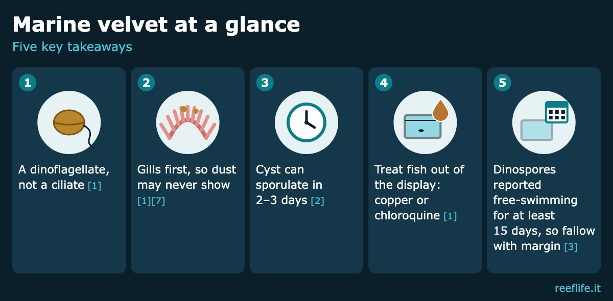
Marine velvet is caused by Amyloodinium ocellatum. It is not a ciliate like ich. It is a parasitic dinoflagellate [1], the same broad group as the zooxanthellae living inside your corals. Here is why marine velvet moves so fast, how to confirm and treat it, and what sources say about fallow.
What is Amyloodinium ocellatum?
Amyloodinium ocellatum is a dinoflagellate ectoparasite. It is the only recognised species in its genus, and it is one of very few organisms that infect both bony fishes and elasmobranchs (sharks and rays) [1]. Its feeding stage is pigmented and photosynthetic, but it lives as a parasite on fish [5]. Merck’s veterinary manual ranks marine velvet among the most serious health problems of captive marine fish; it is also called rust, gold-dust and coral disease [5].
Marine velvet vs ich: not the same enemy
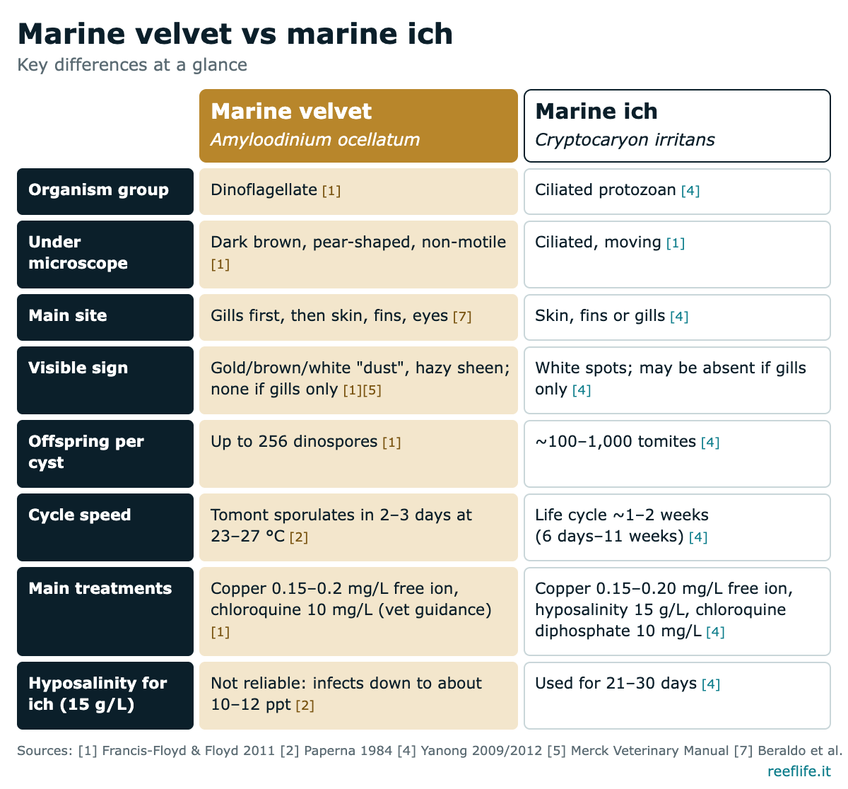
Many keepers meet marine ich (Cryptocaryon irritans) first and assume marine velvet behaves the same way. It does not:
- Organism group. Ich is a ciliated protozoan [4]. Velvet is a dinoflagellate [1].
- Under the microscope. Ich trophonts are ciliated and move. Velvet trophonts are dark brown, ovoid to pear-shaped and do not move [1].
- Main site. In velvet, the gills are the primary site of infection, followed by skin, fins and eyes [7].
- Cycle speed. Ich’s life cycle averages about 1–2 weeks and can range from 6 days to 11 weeks [4]. Under good conditions velvet tomonts can sporulate in 2–3 days [2].
The marine velvet life cycle: three stages
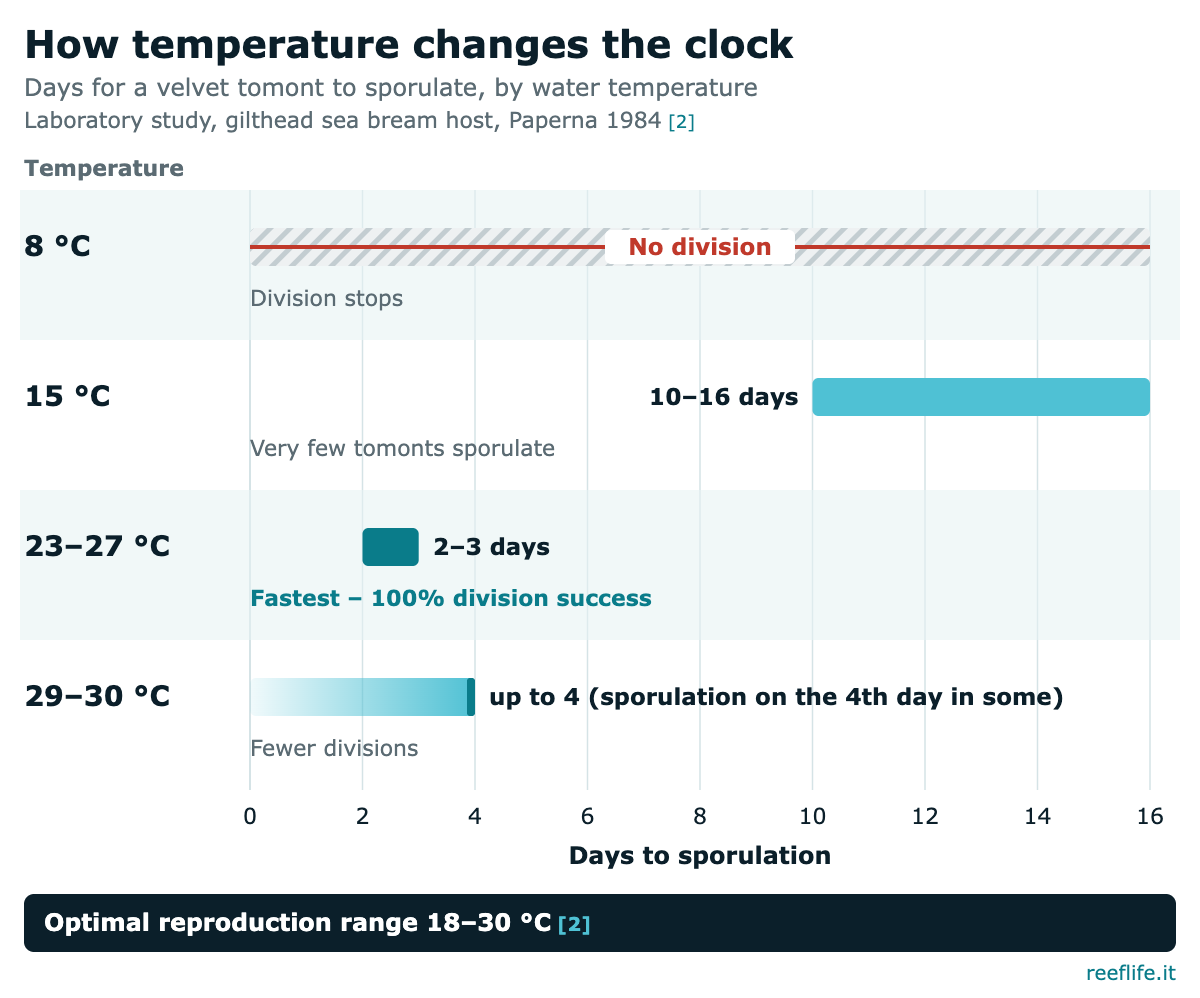
Velvet has a simple, single-host life cycle with three stages [1]:
- Trophont, the feeding stage. It attaches to gills, fins and skin by root-like rhizoids. These penetrate the epithelial cells and cause “degeneration and death of the cell” [8]. At 24 °C trophonts usually stay attached for 2–5 days, although some drop off within 24 hours [1].
- Tomont, the cyst. The mature trophont detaches, settles and divides inside a cyst [1]. A tomont can release up to 256 infective dinospores [1][7].
- Dinospore, the free-swimming infective stage. Dinospores measure 12–15 µm and swim off to find a host [1].
How temperature and salinity change the clock
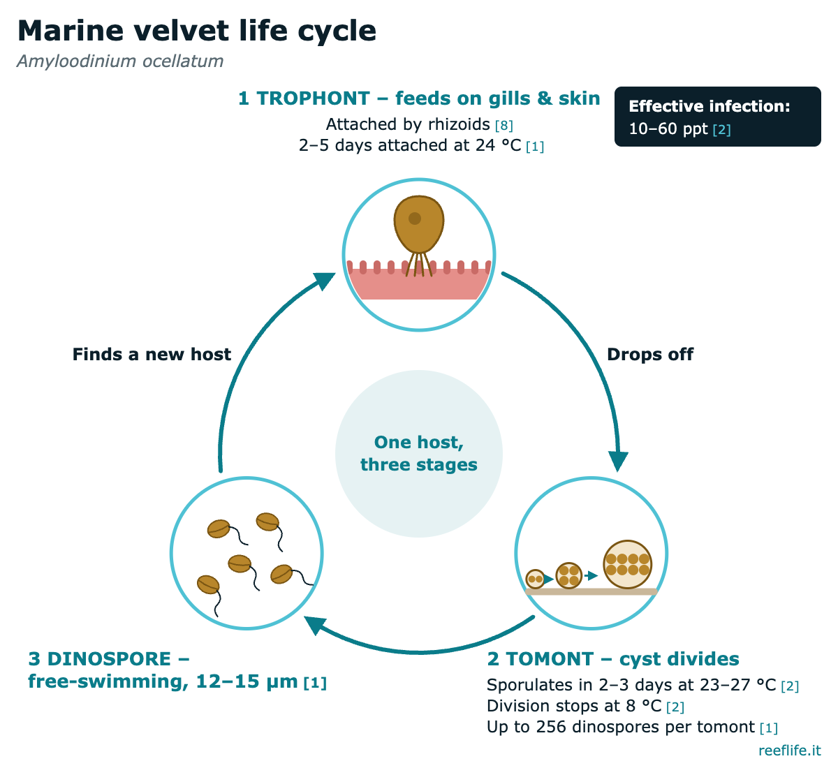
The most detailed laboratory work is Paperna’s 1984 study on gilthead sea bream postlarvae [2]:
- The optimal reproduction range was 18–30 °C. The fastest divisions happened at 23–27 °C, where tomonts took 2–3 days to sporulate [2].
- At 15 °C, very few tomonts sporulated, and only after 10–16 days. At 8 °C, division stopped completely [2].
- At 29–30 °C, the number of divisions declined and some tomonts sporulated only on the 4th day [2].
- Divisions occurred anywhere between 1 and 78 ppt salinity. Full sporulation and effective infection of fish occurred only between 10 and 60 ppt, although the paper’s discussion gives a narrower effective range of 12–50 ppt [2].
Other sources differ: 3–6 days from detachment to dinospore emergence for a Red Sea isolate at 20 °C [1]; a full cycle of 5–7 days [7] or 7–20 days [8], depending on temperature. Typical reef temperatures fall inside the range where marine velvet reproduces fastest, and normal reef salinity (see why salinity matters in a reef aquarium) is well inside its comfort zone [2].
Why marine velvet kills so fast
1. It goes for the gills first. The gills are the primary site of infection [7], and the rhizoids destroy the cells they penetrate [8]. Infected gills may show hyperplasia (tissue overgrowth) and lamellar fusion [1]; heavy infestations add inflammation, haemorrhage and necrosis [6]. Such gills cannot exchange gas or salts properly; researchers list the main causes of death in amyloodiniosis as anoxia, osmoregulatory impairment and secondary microbial infections [6].
2. The visible sign comes late, or never. A gill-only infection does not produce the velvet appearance [1]. In massive infections of European sea bass, fish died “without obvious gross skin alterations” [7]. Affected fish may die suddenly, showing few clinical signs [1].
3. The cycle is short. Each tomont can make up to 256 dinospores [1], while ich cysts actually release more, roughly 100–1,000 tomites [4]. The difference is time: at typical tank temperatures marine velvet can complete reproduction in a few days [2], so the parasite load in a closed system explodes.
How fast is “fast”? In Lawler’s experiments, fish placed in tanks with high dinospore numbers died as early as 12 hours later [3]. In a 2019 challenge trial, white seabream were exposed to about 8,000 dinospores/mL at 24 °C. Mortality reached 100% of the smaller fish and 33.3% of the larger fish within 24 hours [6]. These are heavy experimental doses, not a typical marine velvet outbreak, but they show how little time a gill infection leaves.
Signs and diagnosis of velvet disease
Gill signs: faster gill movement, “piping” and gathering at the surface, reduced appetite [1].
Skin signs: a white or brown “velvet” or hazy, cloudy look that is easiest to see under indirect light, plus flashing, rubbing, poor feeding and weight loss [1]. The colour it gives fish explains its other names, rust and gold-dust [5]. Merck describes small golden spots on the skin that cannot be seen if the infection is only on the gills [5].
Confirming marine velvet. Diagnosis means finding the parasite: a gill or skin biopsy examined as a wet mount at 40× and 100× [1]. Two practical points [1]:
- Prepare wet mounts with seawater of matching salinity. In fresh water the parasite detaches, which can give a false negative.
- Take the sample before anaesthesia, because MS-222 can also cause the parasite to detach.
PCR can detect a single parasite [1], and PCR and rDNA sequencing have been used to confirm outbreaks [9][10].
Treatments: what the evidence supports
None of the common treatment chemicals kills the encysted tomont [8]; only the free-swimming dinospore is susceptible to drugs, so marine velvet treatment must last long enough to catch every wave of dinospores, and extended treatments of more than 10–14 days are needed [1]. Treatment is one of the better-studied aspects of the disease [11], but most evidence comes from aquaculture, not reef fish.
1. Copper (fish-only hospital tank)
Copper is the most commonly used treatment, especially in quarantine systems [1]. The SRAC fact sheet recommends keeping 0.15–0.2 mg/L free copper ion for at least 2–3 weeks. It advises measuring daily, or twice daily if possible, because free copper does not stay in solution and the margin of safety is low [1]. Copper is highly toxic to invertebrates and should not be used where they are housed [1]. Copper is also more toxic to elasmobranchs (sharks and rays) than to bony fish [1]. Use a test kit matched to your copper product and follow its label.
2. Freshwater dip (first aid only)
A 1–3 minute freshwater dip detaches trophonts [1]. A Virginia Tech sheet cites 3–5 minutes to reduce trophonts on gills and skin [8]. A dip may not dislodge all trophonts, and it is unclear whether the detached trophonts are killed [1]; and dips alone are probably not sufficient to stop the parasite spreading [1]. Use it to buy time, never as the marine velvet cure.
3. Chloroquine diphosphate (veterinary guidance)
Chloroquine was rated, along with copper sulfate, among the most effective treatments, at 10 mg/L as an indefinite bath [5]. The SRAC sheet gives 10 mg/L, noting that the system may need to be re-dosed after 7–8 days, with treated water passed through activated carbon before disposal; it is not permitted for food fish [1]. UF/IFAS lists 10 mg/L chloroquine diphosphate as a prolonged bath for 2–3 weeks or more against protozoans including Amyloodinium, and advises checking local regulations before using any drug [4]. Chloroquine is a human medicine with country-specific access rules; use it only under veterinary or fish-health professional guidance.
4. Formalin and hydrogen peroxide (aquaculture tools)
At 25 mg/L, formalin makes trophonts drop off within hours, but it does not kill tomonts [1]. A single 30-minute treatment with 25 mg/L of 35% hydrogen peroxide cut daily losses of mullet fry from 200–1,000 to fewer than 10 within 3 days, though repeat treatments are likely needed [1]. These are aquaculture results, not home-reef first choices.
5. Hyposalinity: don’t import ich advice
Hyposalinity is where marine velvet differs most from ich. For ich, UF/IFAS describes holding salinity at 15 g/L for 21–30 days [4]. Velvet divides between 1 and 78 ppt and infects effectively down to about 10–12 ppt [2]. In a 2025 trial, salinities above 10 ppt supported the parasite’s life cycle [9]. Outbreaks in estuarine fish have occurred at 2–3 ppt [1]. The sources are cautious about how well low salinity works. One says reducing salinity to below 10 ppt for at least 2–3 weeks “will usually be sufficient” for most stenohaline marine fish, though it may be less effective for euryhaline estuarine species [1]. A 2025 trial found that below 5 ppt, 90% of tomonts were degraded within 14 days [9]. Gradually lowering salinity combined with copper worked better than copper alone in farmed sea bass fry [10]. Ich-style hyposalinity (15 g/L) is not a marine velvet treatment. Going below 10 ppt has some support [1][9]. But trophonts on the fish may survive, and the method may fail in euryhaline species [1], so treat it as a tool for professionals.
What this means for your tank
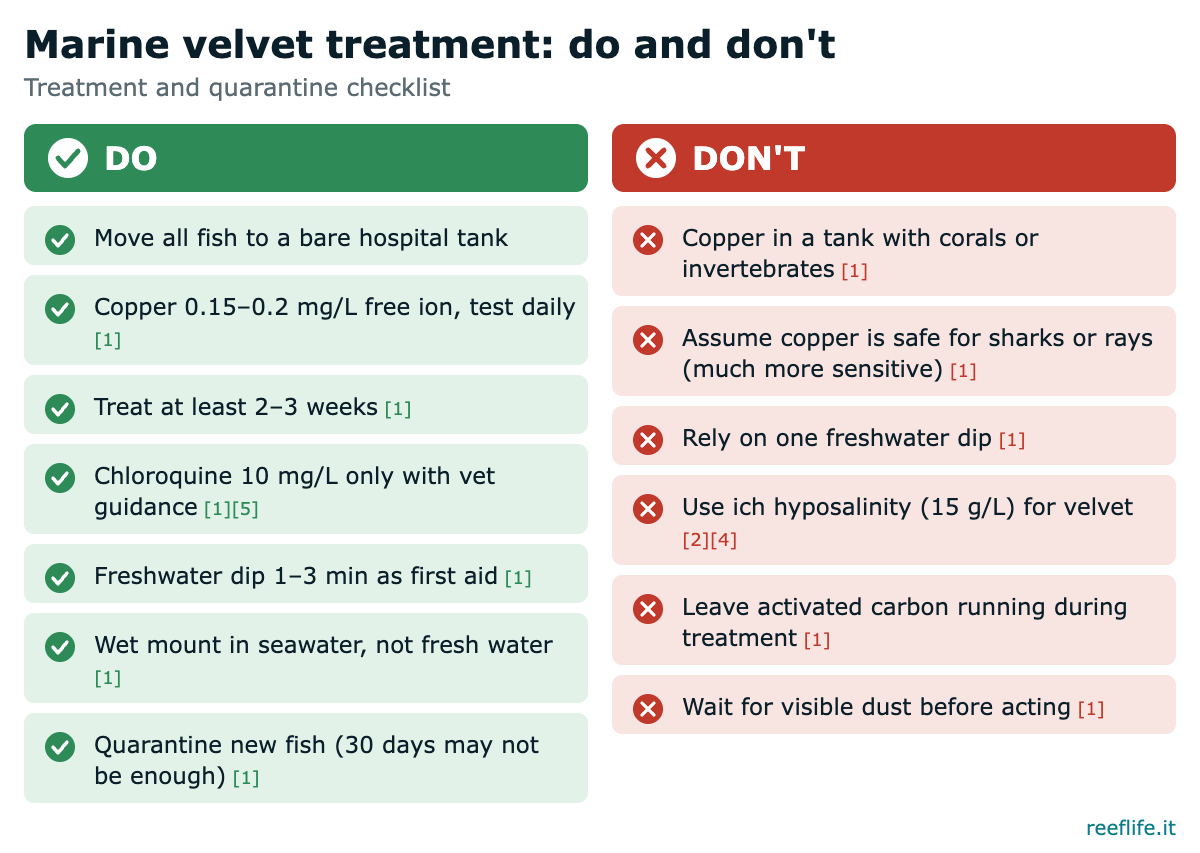
- Act on breathing, not on spots. Rapid gill movement, surface piping and sudden appetite loss across several fish are early warnings [1].
- Confirm if you can. A gill or skin wet mount in tank water separates velvet from ich [1].
- Move every fish to a bare hospital tank. Copper must never be used in a system housing invertebrates [1], so fish are treated away from the display.
- Give a short freshwater dip on transfer, as first aid only [1].
- Start a proven therapy: copper at the stated free-ion range with frequent testing [1], or chloroquine under veterinary guidance [1][5]. Keep activated carbon offline during treatment [1].
- Treat long enough. At least 2–3 weeks for copper [1] and more than 10–14 days overall [1].
- Leave the display without fish. See the fallow section below.
- Quarantine every new fish. A 30-day minimum is often suggested, but it may not be enough [1]. Never add outside water or the water new fish arrive in to your system [1]. Our guide to common marine fish diseases covers the wider quarantine routine.
Fallow period: what the sources really say
Fallow means no fish, so the parasite has no host. None of the scientific sources checked gives a validated marine velvet fallow length for a home display. What they report:
- Dinospore survival. Dinospores remain infective for at least 6 days, but infectivity drops sharply after 24 hours [1]. Lawler, however, kept dinospores free-swimming for at least 15 days [3]. The figures differ widely.
- Dormant tomonts. Tomonts held below 15 °C for 17 weeks still produced infective dinospores after warming, according to an unpublished study cited in the fact sheet [1]. Hibernated tomonts at 16 °C stayed viable for over 6 months [7]. Both come from cool temperatures, but show that the cyst can wait.
A fallow shorter than 15 days would not even cover the longest reported dinospore survival [3]; any longer period is a safety margin, not a proven figure. Corals and invertebrates can stay in the display during fallow. They cannot go into a copper hospital tank [1].
Myths about marine velvet
- “No spots means no disease.” Gill-only infections show no velvet coat [1].
- “It’s just fast ich.” It is a different organism group with a different cycle and different treatment responses [1][4].
- “Hyposalinity fixes everything.” Velvet infects effectively down to about 10–12 ppt [2], well below the 15 g/L used against ich [4].
- “One freshwater dip cures it.” Dips detach many trophonts, but not always all of them, and dips alone are probably not sufficient [1].
- “Copper in the display is fine if I’m careful.” Copper is highly toxic to invertebrates [1].
FAQ
How fast can marine velvet kill a fish?
With very high dinospore numbers, experimental fish died as soon as 12 hours after exposure [3][7]. In most outbreaks, fish weaken over a few days and may die suddenly with few signs [1].
Can I see marine velvet with my eyes?
Sometimes: a gold, brown or white dusty sheen under indirect light, but gill-only infections are invisible [1][5].
Is velvet disease treatable in a reef display?
Not safely with copper, which is highly toxic to invertebrates [1]. Move the fish to a hospital tank.
How long should I quarantine new fish?
The SRAC sheet says a 30-day minimum is often suggested but may not be enough [1].
Summary
Marine velvet is a dinoflagellate parasite, not a ciliate. It attacks the gills first, and its three-stage cycle can turn over in days at reef temperatures [1][2]. That is why fish can die before any velvet appears. Act on fast breathing, confirm with a wet mount, move all fish to a hospital tank, and treat with copper or chloroquine (under professional guidance) for long enough. Leave the display fishless with a generous margin, and never assume ich hyposalinity advice works for marine velvet.
References
- Francis-Floyd R, Floyd MR. Amyloodinium ocellatum, an Important Parasite of Cultured Marine Fish. SRAC Publication No. 4705, Southern Regional Aquaculture Center, 2011. https://srac.tamu.edu/fact-sheets/serve/227
- Paperna I. Reproduction cycle and tolerance to temperature and salinity of Amyloodinium ocellatum (Brown, 1931) (Dinoflagellida). I. Annales de Parasitologie Humaine et Comparée 1984; 59(1): 7–30. https://doi.org/10.1051/parasite/1984591007 (PDF: https://www.parasite-journal.org/articles/parasite/pdf/1984/01/parasite1984591p7.pdf)
- Lawler AR. Studies on Amyloodinium ocellatum (Dinoflagellata) in Mississippi Sound: natural and experimental hosts. Gulf Research Reports 1980; 6(4): 403–413. https://doi.org/10.18785/grr.0604.08
- Yanong RPE. Cryptocaryon irritans Infections (Marine White Spot Disease) in Fish. UF/IFAS EDIS FA164, 2009 (revised 2012). https://ask.ifas.ufl.edu/publication/FA164/pdf
- Petty BD, Francis-Floyd R, Yanong RPE. Parasitic Diseases of Fish. Merck Veterinary Manual, Professional Version (reviewed 2022). https://www.merckvetmanual.com/exotic-and-laboratory-animals/aquarium-fishes/parasitic-diseases-of-fish
- Moreira M, Cordeiro-Silva A, Barata M, Pousão-Ferreira P, Soares F. Influence of age on stress responses of white seabream to amyloodiniosis. Fishes 2019; 4(2): 26. https://doi.org/10.3390/fishes4020026
- Beraldo P, Massimo M, Galeotti M. Amyloodinium ocellatum. In: Sitjà-Bobadilla A et al. (eds), Fish Parasites, CABI, 2024 (chapter 4, Open Access; chapter copyright line: © 2021). https://doi.org/10.1079/9781800629127.0004 (PDF: https://air.uniud.it/retrieve/ec9de723-6bf3-4dae-8fdc-c033c928e05f/beraldo-et-al-2024-amyloodinium-ocellatum.pdf)
- Smith SA, Schwarz MH. Getting Acquainted with Amyloodinium ocellatum. Virginia Cooperative Extension Publication 600-200, 2019. https://ext.vt.edu/content/dam/pubs_ext_vt_edu/600/600-200/CNRE-39P.pdf
- Vidya R, Ananda Raja R, Avunje S, et al. A report on outbreak of Amyloodinium ocellatum infestation in broodstock of Java rabbitfish, Siganus javus (Linnaeus, 1766). Journal of Parasitic Diseases 2025; 49(1): 45–56. https://doi.org/10.1007/s12639-024-01710-1
- Bessat M, Fadel A. Amyloodiniosis in cultured Dicentrarchus labrax: parasitological and molecular diagnosis, and an improved treatment protocol. Diseases of Aquatic Organisms 2018; 129: 41–51. https://doi.org/10.3354/dao03237
- Moreira M, Costas B, Rodrigues P, et al. Amyloodiniosis in aquaculture: a review. Reviews in Aquaculture 2023. https://doi.org/10.1111/raq.12883
About the author
AVen is a reef keeper and the author of reeflife.it, a blog about keeping marine reef aquariums. Read more about AVen and reeflife.it.


About Author
